Anatomy Pictures Of Lower Back And Hip - Hip Pain Explained Including Structures Anatomy Of The Hip And Pelvis - These sections are cervical (neck), thoracic (upper and middle back), lumbar (lower back), and sacrum (tailbone).
Anatomy Pictures Of Lower Back And Hip - Hip Pain Explained Including Structures Anatomy Of The Hip And Pelvis - These sections are cervical (neck), thoracic (upper and middle back), lumbar (lower back), and sacrum (tailbone).. Collection by remove back pain for good. Anatomy pictures of lower back and hip : Related online courses on physioplus. You can move going back and forth in the fashion that i am right now, in many of you will have this muscle be really, really tight because your gluteus from the lower aspects of the gluteus maximus, those areas are not firing very well. A ct scan using contrast dye can also provide a useful picture of the spinal.
Related online courses on physioplus. Hip anatomy, function and common problems. Collection by remove back pain for good. Your lower back (lumbar spine) is the anatomic region between your lowest rib and the upper part of the these nerves also control movements of your hip and knee muscles. By dr arun pal singh.
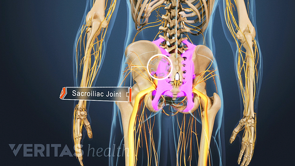
Types Of Arthritis That Cause Sacroiliac Joint Pain from embed.widencdn.net The sacrum and hip bones form a ring called the pelvic girdle. The hip joint is one of the most flexible joints in the entire human body. The iliopsoas muscle, which extends from the lower back to. Place your hand under the lumbar spine to detect masking of restricted hip joint. Want to learn more about it? Left superficial lymphatic vessels of back. A collection of anatomy notes covering the key anatomy concepts that medical students need to learn. Anatomy pictures of lower back and hip :
This arrangement gives the hip anatomy a large amount of motion needed for daily activities.
The most common symptom of sciatica is lower back pain that extends through the hip and buttock and down one leg. Left superficial lymphatic vessels of back. —the hip bones are largely covered with muscles, so that only at a few points do they approach the surface. Related online courses on physioplus. Hip anatomy, function and common problems. Picture a man standing with the front of his pelvis tilting forward and his tailbone lifting. Knowing the anatomy of your hip can help you understand the source of any hip pain. These sections are cervical (neck), thoracic (upper and middle back), lumbar (lower back), and sacrum (tailbone). A ct scan using contrast dye can also provide a useful picture of the spinal. Understanding how the different layers of the hip are built and connected can help you understand how the hip works, how it can be injured, and how challenging recovery can be when this joint is injured. The foot swings forward and comes back into contact with the floor with a heel strike active hip flexion. Learn about anatomy lower limb with free interactive flashcards. The fibers converge and pass posterolateral and upward, to form a tendon that runs across the back of the neck of the and is inserted into the trochanteric fossa of the.
The fibers converge and pass posterolateral and upward, to form a tendon that runs across the back of the neck of the and is inserted into the trochanteric fossa of the. In vertebrate anatomy, hip (or coxa in medical terminology) refers to either an anatomical region or a joint. The most common symptom of sciatica is lower back pain that extends through the hip and buttock and down one leg. Read about associated symptoms and signs, and learn about diagnosis, prognosis, treatment, and the types of specialists who treat hip pain. On anatomical parts the user can choose to display the bones (pelvis, femur, tibia, fibula, patella, foot bones) and the different joints (hip joint, femorotibial joint, ankle joints and foot.
How To Deal With A Pinched Nerve In Your Hip With Pictures from www.wikihow.com Picture a man standing with the front of his pelvis tilting forward and his tailbone lifting. A collection of anatomy notes covering the key anatomy concepts that medical students need to learn. Collection by remove back pain for good. When a person experiences lower back and hip pain simultaneously, there may be an underlying injury or medical condition causing both of these symptoms. Learn about anatomy lower limb with free interactive flashcards. The hip muscles are going to be slip into hip muscles and gluteal muscles. When most people mention their back, what they are actually referring to is their spine. Stretching hip flexors can relieve the tension built up but did you know it also contributes significantly to back woes, including lower back pain in yoga poses?
Place your hand under the lumbar spine to detect masking of restricted hip joint.
Understanding how the different layers of the hip are built and connected can help you understand how the hip works, how it can be injured, and how challenging recovery can be when this joint is injured. Possible causes of lower back and hip pain include sprains, strains, and a herniated disk. So because of that your glute medius tends to have to has to has. Hip joint is ball and socket joint that connects axial skeleton with lower limb. The hip muscles are going to be slip into hip muscles and gluteal muscles. By dr arun pal singh. Five vertebrae (l1 to l5) make up the lower part of the spine. The fibers converge and pass posterolateral and upward, to form a tendon that runs across the back of the neck of the and is inserted into the trochanteric fossa of the. Four fused vertebrae this part of your anatomy is susceptible to injury, arthritis, herniated disks, pinched nerves and other problems. Left superficial lymphatic vessels of back. When a person experiences lower back and hip pain simultaneously, there may be an underlying injury or medical condition causing both of these symptoms. The human spine is composed of 4 sections of vertebrae. I had coccyx bend.i had injection, rectification, and it just.
Continue scrolling to read more below. Anatomy pictures of lower back and hip : The foot swings forward and comes back into contact with the floor with a heel strike active hip flexion. Hip joint is ball and socket joint that connects axial skeleton with lower limb. The iliopsoas muscle, which extends from the lower back to.
Lower Extremity Anatomy Bones Muscles Nerves Vessels Kenhub from thumbor.kenhub.com When a person experiences lower back and hip pain simultaneously, there may be an underlying injury or medical condition causing both of these symptoms. I had coccyx bend.i had injection, rectification, and it just. Picture a man standing with the front of his pelvis tilting forward and his tailbone lifting. In vertebrate anatomy, hip (or coxa in medical terminology) refers to either an anatomical region or a joint. The hip muscles are going to be slip into hip muscles and gluteal muscles. These sections are cervical (neck), thoracic (upper and middle back), lumbar (lower back), and sacrum (tailbone). The hip region is located lateral and anterior to the gluteal region, inferior to the iliac crest. On anatomical parts the user can choose to display the bones (pelvis, femur, tibia, fibula, patella, foot bones) and the different joints (hip joint, femorotibial joint, ankle joints and foot.
Understanding how the different layers of the hip are built and connected can help you understand how the hip works, how it can be injured, and how challenging recovery can be when this joint is injured.
So because of that your glute medius tends to have to has to has. Your lower back (lumbar spine) is the anatomic region between your lowest rib and the upper part of the these nerves also control movements of your hip and knee muscles. The sacrum and hip bones form a ring called the pelvic girdle. Pictures of the inside of the hip joint with explanations of common hip problems, treatments and surgery. By dr arun pal singh. Possible causes of lower back and hip pain include sprains, strains, and a herniated disk. Knowing the anatomy of your hip can help you understand the source of any hip pain. While the thigh muscles will be slip into the anterior, medial and posterior groups. When most people mention their back, what they are actually referring to … The most common symptom of sciatica is lower back pain that extends through the hip and buttock and down one leg. These sections are cervical (neck), thoracic (upper and middle back), lumbar (lower back), and sacrum (tailbone). The pain usually affects only an mri can show the alignment of vertebral disks, ligaments, and muscles. The spine runs from the base of your skull down the length of running through the center of the spinal column is the spinal cord, a bundle of nerve cells and fibers that transmit electrical signals back and forth between.

No comments:
Post a Comment